 Image credit:Figure2
Abstract
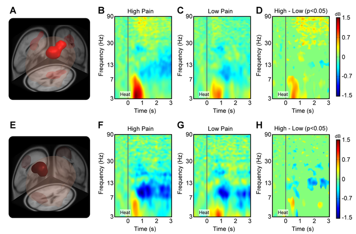
The translation of brief, millisecond-long pain-eliciting stimuli to the subjective perception of pain is associated with changes in theta, alpha, beta, and gamma oscillations over sensorimotor cortex. However, when a pain-eliciting stimulus continues for minutes, regions beyond the sensorimotor cortex, such as the prefrontal cortex, are also engaged. Abnormalities in prefrontal cortex have been associated with chronic pain states, but conventional, millisecond-long EEG paradigms do not engage prefrontal regions. In the current study, we collected high-density EEG data during an experimental paradigm in which subjects experienced a 4-s, low- or high-intensity pain-eliciting stimulus. EEG data were analyzed using independent component analyses, EEG source localization analyses, and measure projection analyses. We report three novel findings. First, an increase in pain perception was associated with an increase in gamma and theta power in a cortical region that included medial prefrontal cortex. Second, a decrease in lower beta power was associated with an increase in pain perception in a cortical region that included the contralateral sensorimotor cortex. Third, we used machine learning for automated classification of EEG data into low- and high-pain classes. Theta and gamma power in the medial prefrontal region and lower beta power in the contralateral sensorimotor region served as features for classification. We found a leave-one-out cross-validation accuracy of 89.58%. The development of biological markers for pain states continues to gain traction in the literature, and our findings provide new information that advances this body of work.